
Leica DMi8
https://labs.jhu.edu/wp-content/uploads/2016/01/User-Manual-DMi8.pdf
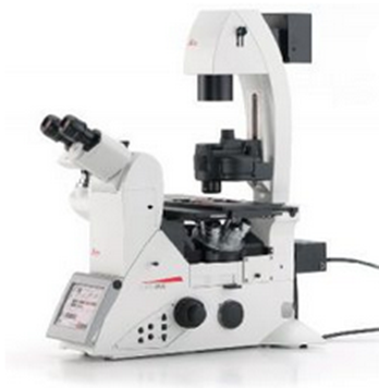
The Leica DMi8 is a state-of-the-art automated microscope that provides a comprehensive solution for a wide range of applications and enables researchers to streamline their workflow and increase their efficiency.
- Automated Focus: The automated focus function allows the microscope to automatically adjust the focus on the sample, saving you time and minimizing potential errors that may occur with manual adjustments.
- Z-Stacking: Z-stacking allows the microscope to automatically capture a series of images at different focus planes, creating a 3D image of the sample. This is especially useful when examining samples that lie deeper within the sample.
- Live Image Capture: Live image capture allows you to observe the sample in real-time on your computer screen without having to take a new image each time.
- Multi-Channel Fluorescence Support: The DMi8 supports multi-channel fluorescence, allowing you to examine multiple fluorescent dyes in a sample simultaneously.
- Time-Lapse Capture: With time-lapse capture, you can set the microscope to automatically capture images at specific times. This is particularly useful for longer studies where you may want to observe the change in a cell over time.
- Customizable Grid Scan: The DMi8 allows you to create your own custom grid scans, which can then be automatically traversed, taking images at the programmed intervals.
- Integrated Illumination Modes: The DMi8 has a range of integrated illumination modes, including brightfield, fluorescence, and phase contrast, providing you with a variety of observation options.